With the gradual increase in demand for healthcare services, people need to be able to treat diseases as well as minimize wound scars. As a result, the need for cosmetic surgical suturing techniques is also gradually increasing
Anatomical Structure of The Skin
The skin consists of two layers: the epidermis and the dermis. The epidermis consists of the stratum corneum (upper layer) and the non-corneal layer (lower layer). The color of the skin deepens with an increase in the content of melanocytes, and vice versa the color of the skin becomes lighter.
The dermis is generally divided into papillary and reticular layers, which are directly nourished by blood vessels and nerves, and are rich in elastic and collagen fibers that are responsible for the skin’s elasticity and its ability to contract.
Skin Relaxation Tension Lines

The wrinkle lines are perpendicular to the direction of the muscle fibers, whereas the RSTL refers to the point in the skin where the collagen, elastic fibers, etc. form the least amount of tension. The RSTL corresponds to the spontaneous formation of wrinkles after skin relaxation, and is therefore approximately the same, but differs in some areas, such as between the eyebrows and the side of the nose.
When the incision corresponds to the direction of the RSTL, a wound with less tension is obtained, which leads to the fastest healing and the least scar formation; when there is a crease, the incision should be designed within the skin fold, following the principle of “hiding the scar within the skin fold”.
Principles of Cosmetic Skin Suturing Techniques
The wound should be slightly flared after skin suturing, and the suture should be taut enough but not too tight, while the knot needs to be buried deep in the tissue to minimize irritation of the knot and avoid scarring.
The Hollander Wound Evaluation Scale (HWES) suggests that ideal wound healing should meet the following six key points:
1, No tissue misalignment;
2, Neat wound alignment;
3, Wound alignment spacing of no more than 2mm;
4, No skin edge inversion;
5, No excessive distortion of skin tissue;
6, Overall aesthetics.
Achievement of each of the above key points is credited 1 point, 6 is the best wound healing.
Standard Surgical Suturing Techniques
Skin wound closure usually consists of subcutaneous and cutaneous sutures. Subcutaneous sutures are generally used to reduce tension on both sides of the wound using interrupted sutures and allow the knot to be buried deeper in the tissue.
It is important to note that the subcutaneous tissue is less resilient and does not provide adequate reduction of tension; therefore , a small amount of dermal tissue may be carried in the subcutaneous suture. In addition, subcutaneous adipose tissue can be trimmed prior to suturing to prevent tissue buildup from interfering with the alignment of the skin margins and insertion between the skin margins to interfere with healing.
Skin sutures are used to re-form the skin edge using interrupted sutures. It is generally recognized that interrupted sutures are the most commonly used and most effective suture for cosmetic dermatologic closure because of the ease of regulating local tension and the fact that adverse events such as poor suture results and loose knots in a single site have less impact on the overall appearance.
However, it should be noted that interrupted sutures require more knots and more sutures, so the surgeon will spend more time and patience.
Irregular Wound Suturing Techniques
- The difference in length between the two sides of the wound is less than 1cm, the middle of the wound is sutured first, followed by the middle of both sides of the wound, and then the middle of each individual wound continues to be sutured until the wound is completely closed.
2、When the difference in length between the two sides of the wound is less than 1cm, in order to avoid the appearance of small “cat ears” at both ends , you can also be sutured at both ends to confirm the flatness of the wound, and then follow the technique 1 to close the wound.
- If the difference in length between the two sides of the wound is greater than 1 cm, “cat ears” will inevitably appear. In this case, suturing is started from one end of the wound, and the excess skin on the longer side is “driven” to one end. Subsequently, an oblique incision (1-2cm) is made, and the excess skin outside the incision is excised and sutured.
Rules and Tips – Proper Surgical Suturing Techniques Results Depend on:
(1) The wound edges of the skin must be of similar length, otherwise “cat ears” will form. The latter can be eliminated by trimming or other techniques.
(2) Wound edges with depth can be brought to the same level by skin excision and skin movement.
(3) The entrance and exit depths of the subcutaneous suture area must be equal (otherwise deformation of the wound edge will result).
(4) The knot of the skin suture must not be pulled too tightly, otherwise scar constriction will develop (postoperative wound swelling must be taken into account).
(5) The ends of the sutures must be left long enough for removal but must be cut short enough to prevent them from interfering with adjacent sutures.
(6) After the suture is completed, the wound edges should be inspected. The epithelium should not be involved but should be turned outward.
- Circular irregular wounds.
(1) If the short and long axes of the wounds are equal, the wounds can be trimmed to a shuttle shape and sutured along the line of skin relaxation tension or perpendicular to the direction of greater skin tension.
(2) If the short and long axes of the wounds are more different, trim the wounds in the direction of the long axis and close them in the shape of a shuttle. It should be noted that the initial trimming of the wound to avoid the removal of a large amount of skin tissue, can be in the suture alignment according to the treatment of linear irregular wounds and then trimming.
- Triangular-like irregular wound.
(1) When the triangle is an obtuse triangle or when there is little tension in one direction of the triangle, the wound can be trimmed to an obtuse triangle and sutured to a linear shape. Localized accumulations of skin tissue are reprocessed in the same manner as for linear irregular wounds.
(2) If the triangle cannot be trimmed or the wound tends to be triangular, the wound can be treated with a triangular suture, i.e., the tips of the three sides are sutured together and the rest of the wound is treated as a linear suture. When suturing the tips of the three sides, carry the tissue of the tip of side b after entering the needle from the epidermis of side a (note that tip necrosis may occur when carrying less tissue), and then tie the knot after exiting the needle from the epidermis of side c.
Rules and Tips for Surgical Suturing Techniques:
When closing irregular wounds, try to adjust the direction of the suture to the direction with the least tension on the skin.
Avoid removing too much skin tissue when trimming “cat’s ears”, smaller “cat’s ears” can be improved by themselves.
- Avoid using the suture method shown in the picture, as it may cause ischemic necrosis of the tip and poor healing of the triangular suture.
Special Surgical Suturing Techniques
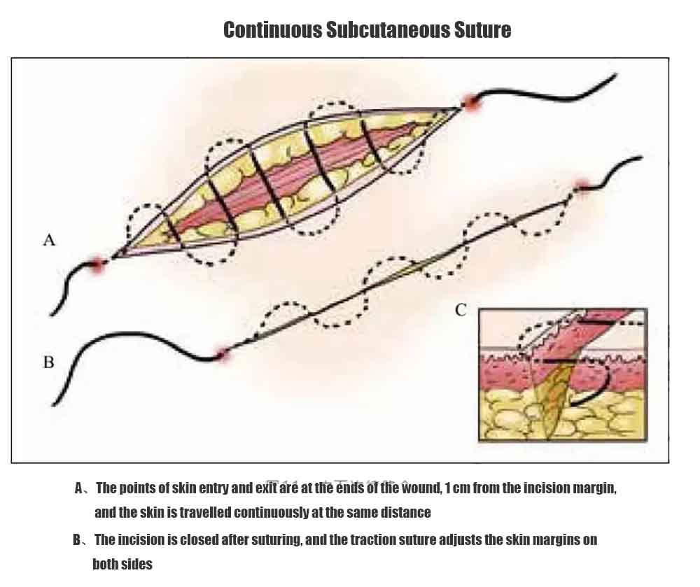
(a) Continuous Intradermal Suture
1、Advantages: The advantage of this suturing technique is that it usually requires only one entrance hole and one exit hole. This avoids the epithelialization of too many puncture holes, especially in areas of the skin rich in sebaceous glands, and is beneficial in reducing postoperative scar formation.
2、Suture method: The suture first enters the skin near one end of the wound and enters the wound intradermally. The suture is then passed through the other side of the wound in the dermal plane at exactly the same length to reach the distal end. The suture then leaves the skin at the distal end of the wound. The length of the wound edge is adjusted by applying slight traction to the suture, and finally, the suture is secured with “buttons”, sterile surgical tape, etc. to prevent inadvertent removal.
Rules and Techniques for Surgical Suturing Techniques:
Continuous subcutaneous sutures are only suitable for incisions with similar lengths of skin edges on both sides:
- It is not well suited for suturing curved wounds, which can lead to distortion of the wound.
- Barbed sutures can be applied to improve suture stability.
(b) Vertical Mattress Suturing (Donati suture)
1, Advantages: Vertical decubitus suture has the advantage of safer reconstruction of incisions with large differences in depth, which makes the skin edge flatter on both sides, and helps to avoid the formation of sulcus scars, or even epithelial inversion, which can lead to poor wound healing.
2, Suture method: The needle entry point is located at about 4mm from the edge of the incision, and then the needle comes out at the same distance from the skin edge on the opposite side, and then the needle comes in at about 1mm from the edge of the incision and comes out at the same distance from the opposite side. The knot can be slightly tightened to flare the incision edge.
Rules and Techniques:
As an alternative a semi-buried vertical mattress suture may also be used. Horizontal mattress sutures also allow flaring of the skin margin but are not as stable as vertical mattress sutures. However, the mattress suture produces four suture marks with each stitch and can affect the hemodynamics of the skin margin, so it should be used with extreme caution.
Q: Are you a factory?
A: Yes, we have more than 25 years of industry experience leading companies
Q: What’s your delivery time?
A: Commonly used products we have sufficient stock, so it usually takes 25-35 days for your order to reach you after receiving your advance payment. This time varies with the instrument type and their quantity which can be negotiated.
Q: What’s the quality of your products?
A: The quality of our surgical tools has gained a superb reputation in the industry. We offer free of cost samples so that our customers can test the quality and precision of the instrument. (NOTE: The customer has to pay shipping/courier charges. )
Q: What services can we provide?
A: We provide excellent after-sale services. All packages/shipments are thoroughly checked, any damaged or defected instruments are replaced before shipment.
Q: Do you support customization?
A: Sure. Customization is welcome!

 Capsulorhexis Forceps
 Suture Forceps
 Hemostatic Forceps
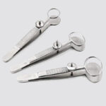 Chalazion Forceps
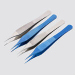 Adson Forceps
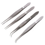 Tissue Forceps


